views

- Patient from Florida Presents with Dermatologic Lesion: Diagnosis for Frequent Sun Tan Enthusiast
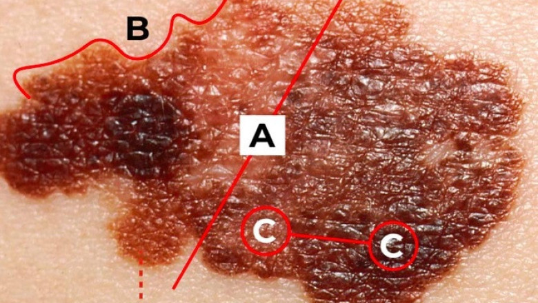
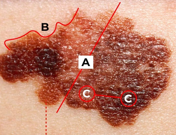
A patient hailing from Florida presents at the healthcare facility with a conspicuous dermatologic lesion. The patient has a noted history of frequent sun tanning and prolonged exposure to ultraviolet radiation. The lesion, a rough and scaly patch, is located on areas commonly exposed to the sun, such as the face, neck, or arms. The patient reports occasional pruritus and discomfort in the affected area.
Clinical Presentation
The primary manifestation of the patient is:
-
Dermatologic Lesion: The patient presents with a rough, scaly patch on sun-exposed skin. The lesion is slightly elevated and has a scaly surface, which is frequently associated with chronic sun exposure.
Differential Diagnosis
Given the patient's predilection for sun tanning and the appearance of the dermatologic lesion, several potential diagnoses should be considered:
-
Actinic Keratosis: This is a precancerous skin condition precipitated by chronic sun exposure. It manifests as rough, scaly patches on sun-damaged skin and has the potential to progress to squamous cell carcinoma.
-
Solar Lentigines: Also referred to as age spots or liver spots, these are flat, hyperpigmented macules that develop on sun-exposed areas of the skin due to cumulative ultraviolet radiation exposure.
-
Basal Cell Carcinoma: This is the most prevalent form of skin cancer, often presenting as a pearly or waxy nodule, or a flat, flesh-colored lesion that may ulcerate or form a crust.
-
Squamous Cell Carcinoma: This type of skin cancer can present as a rough, scaly patch or a firm erythematous nodule, commonly occurring on sun-exposed skin.
-
Seborrheic Keratosis: These benign skin growths appear as rough, waxy, or slightly elevated lesions and are often non-cancerous.
Diagnostic Workup
To determine the underlying etiology of the dermatologic lesion, the following diagnostic tests and evaluations are recommended:
-
Dermatologic Biopsy: A specimen of the lesion can be obtained for histopathological examination to ascertain the exact nature of the cells.
-
Dermoscopy: A non-invasive technique that employs a dermatoscope to examine the lesion's surface and vascular patterns in detail.
-
Serological Testing: To rule out systemic conditions that may contribute to cutaneous changes.
-
Patch Testing: If allergic contact dermatitis is suspected, patch testing can aid in identifying potential allergens.
Conclusion
The patient's presentation with a dermatologic lesion and a history of frequent sun tanning necessitates a comprehensive diagnostic workup to ascertain the precise cause. Early diagnosis and appropriate therapeutic intervention are crucial for managing the condition and averting potential complications. Regular follow-up and monitoring are indispensable to ensure the patient's dermatologic health and overall well-being.


Comments
0 comment